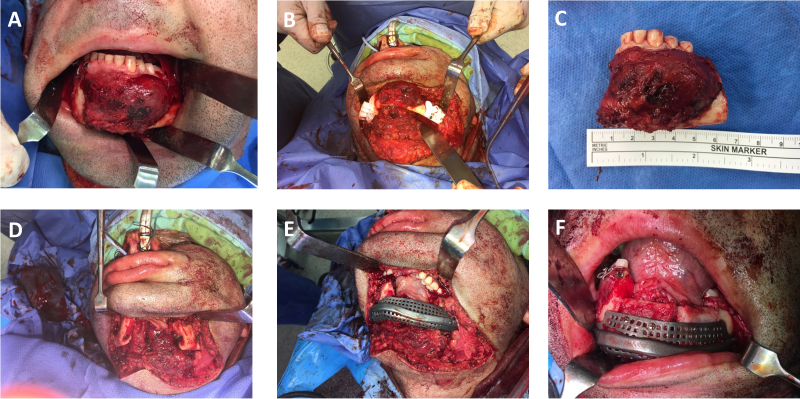
Figure 10. The Operation of the Patient in Figure 9 Is Presented.
A: Tumor exposure. B: Cutting guides are placed. C and D: Following resection. E: Insertion of the patient-specific implants without the need for external fixator as the bony relations are re-established by the drilled holes performed earlier using the cutting guides. F: Placement of the bone graft in the meshed crib.

