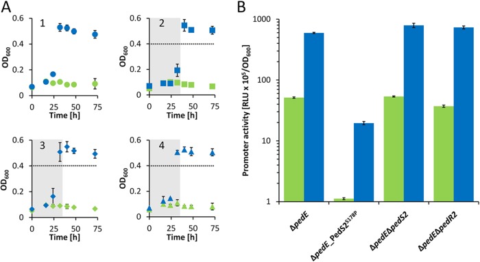
FIG 3 .
(A) Growth of ΔpedE, ΔpedE_PedS2S178P, ΔpedE ΔpedS2, and ΔpedE ΔpedR2 strains. The ΔpedE (circles; panel 1), ΔpedE_PedS2S178P (squares; panel 2), ΔpedE ΔpedS2 (diamonds; panel 3), and ΔpedE ΔpedR2 (triangles; panel 4) were grown at 30°C and 350 rpm shaking with M9 medium in 96-well plates supplemented with 5 mM 2-phenylethanol in the presence of 10 µM La3+ (blue symbols) or absence of La3+ (green symbols). The gray areas in panels 2 to 4 show the time point by which the parental ΔpedE strain (circles) reached their maximum OD600. (B) Activities of the pedH promoter in ΔpedE, ΔpedE_PedS2S178P, ΔpedE ΔpedS2, and ΔpedE ΔpedR2 strains in the presence of 1 µM La3+ (blue bars) or in the absence of La3+ (green bars) or measured in M9 medium supplemented with 1 mM 2-phenylethanol. Promoter activities are presented in relative light units (RLU × 105) normalized to OD600. All data represent the means for biological triplicates, and error bars correspond to the respective standard deviations.

