. 2018 Jul 16;9(33):6758–6764. doi: 10.1039/c8sc02578a

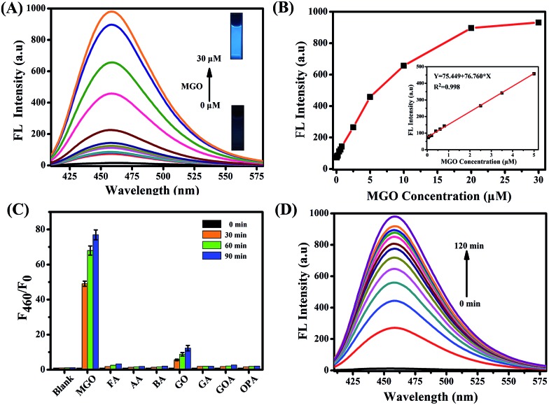
Fig. 1. (A) The fluorescence spectra of NI-OPD (10 μM) with different concentrations of MGO (0–30 μM). (B) The fluorescence change and linear response of NI-OPD (10 μM) to MGO. (C) The fluorescence histogram response of NI-OPD (10 μM) towards various aldehydes including formaldehyde (FA), acetaldehyde (AA), benzaldehyde (BA), glyoxal (GO), glutaraldehyde (GA), glyoxylic acid (GOA), and o-phthalaldehyde (OPA). (D) Time dependent fluorescence intensity changes at 460 nm against the reaction time (0–120 min) of NI-OPD (10 μM) toward MGO (30 μM). All analytes were evaluated at 100 μM, except MGO at 30 μM and the data were obtained in PBS buffer (10 mM, pH = 7.4, 10% DMF). λex = 380 nm.

Fig. 1