. 2018 Jul 13;9(31):6480–6489. doi: 10.1039/c8sc01680a

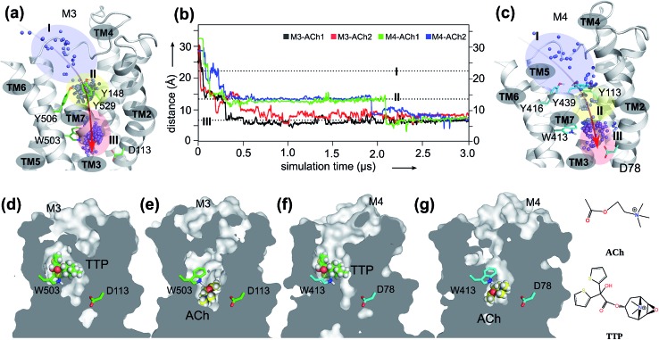
Fig. 1. The orthosteric and novel ligand binding sites of the muscarinic M3 and M4 receptors: (a) view of the M3 receptor showing the overall movement (red arrow) of the ACh molecule from the extracellular vestibule towards the orthosteric site of the receptor during the 3.0 μs MD simulations. Blue dots: mass centers of ACh at different simulation times showing its positional fluctuations within and between the extracellular vestibule, and orthosteric and novel ligand binding sites. Blue-shaded area: extracellular vestibule (zone I). Yellow-shaded area: orthosteric site (zone II). Red-shaded area: novel ligand site (zone III). (b) Time dependence of distance between ACh and oxygen at the sidechain of D2.50 during two independent MD simulations for M3–ACh (black and red) and for M4–ACh (green and blue). The simulations reveal the atomistic details of ACh movements from the extracellular vestibule to the novel ligand binding site of both the M3 and the M4 receptors. (c) View of the M4 receptor showing the overall movement (red arrow) of the ACh molecule from the extracellular vestibule towards the orthosteric site of the receptor during the 3.0 μs MD simulations. Details as in A. (d–g) Cross-sections through the centers of M3 and M4 receptors revealing the final positions of the indicated ligands in different receptor binding sites.

Fig. 1