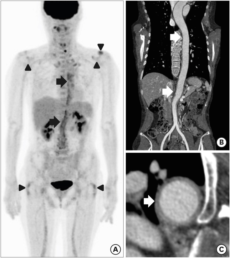
Fig. 1. 18F FDG-PET/CT and CT angiography findings of a 56-year-old female diagnosed with GCA. Coronal FDG-PET/CT (A) image demonstrates a significant uptake in aortic wall from the level of thoracic spine to aortic bifurcation (black arrows). Uptake of the tracer of the bilateral glenohumeral joints, bilateral greater trochanter area and left acromio-clavicle joint is also seen (black arrowheads). CT angiography image (B) and (C) shows the diffuse thickening with enhancement in thoracic aorta and abdominal aorta (white arrows).
FDG-PET/CT = fluorodeoxyglucose positron emission tomography/computed tomography, CT = computed tomography, GCA = giant cell arteritis.

