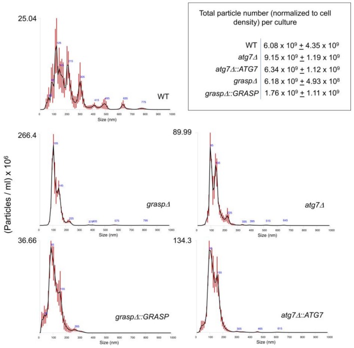
Figure 1.
Nanoparticle tracking analysis of Cryptococcus neoformans extracellular vesicles (EVs) comparing wild type (WT), mutant (graspΔ and atg7Δ) and complemented (graspΔ::GRASP and atg7Δ::ATG7) cells. Results are representative of two independent biological replicates producing similar profiles. Particles were quantified in EV samples suspended in 150 mL phosphate-buffered saline (PBS). Particle detection values shown in the upper, right panel were normalized to the total number of cells in the cultures from which each sample was obtained.

