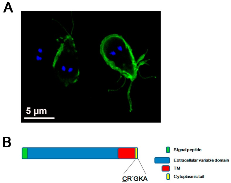
Figure 1.
Variant-specific surface protein or VSP. (A) Confocal immunofluorescence image showing the plasma membrane localization of the VSP9B10 (green) by using FITC-labeled anti-9B10 mAb. The nuclei are stained with DAPI (blue). Scale bars: 5 μm. VSP9B10 covers two of the trophozoites observed. (B) Schematic representation of a VSP containing a signal peptide, an extracellular variable domain, a transmembrane (TM) domain, and the CRGKA invariable cytoplasmic tail. The sites of palmitoylation (underlined C) and citrullination (the asterisk in R) of the cytoplasmic tail are denoted.

