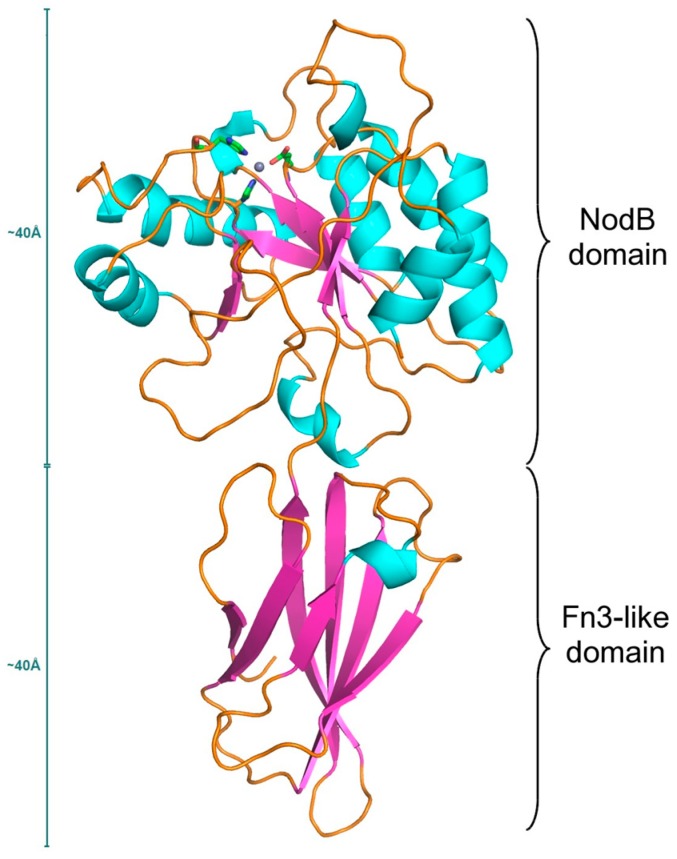
Figure 1.
Ribbon diagram of the Ba0331 polysaccharide deacetylase (PDA) protein architecture showing the two distinct domains. Helices are represented as cyan ribbons, β-strands are in magenta and loops are shown as orange strings. The Zn ion is shown as a grey sphere with the coordinating amino acid residues (Asp213, His271 and His275) in stick representation.

