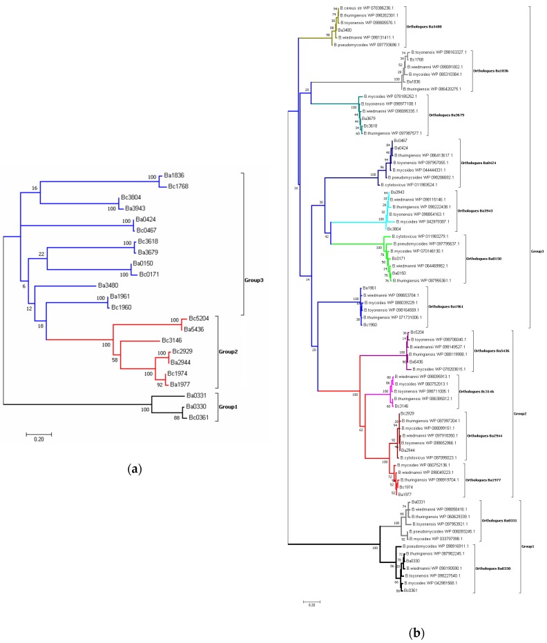
Figure 2.
Molecular phylogenetic analysis of the PDA NodB domain. (a). The unrooted tree of 23 protein sequences of the PDA NodB domain from B. anthracis strain Ames (Ba) and B. cereus strain ATCC14579 (Bc) by the Maximum Likelihood method and with the highest log likelihood (−4084.19) is shown. The sequences are clustered in three groups represented with different colours (Group 1 in black, Group 2 in red and Group 3 in blue). (b). The unrooted tree of the NodB domain of 80 PDA amino acid sequences (homologues of the B. anthracis strain Ames (Ba) and B. cereus strain ATCC14579 (Bc) given in Figure 1a), selected from six different Bacilii species by the Maximum Likelihood method and with the highest log likelihood (−5578.53) is shown. The percentage of trees where the associated taxa clustered together is shown next to the branches. Grouping of the orthologues with respect to the B. anthracis/B. cereus species is shown in different colours.

