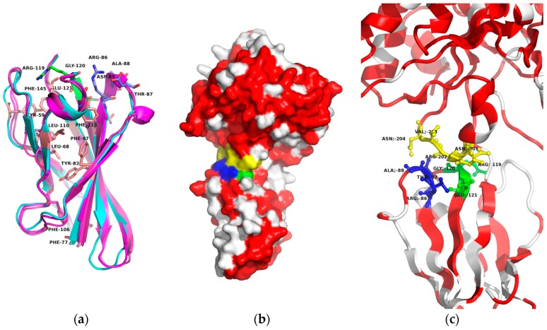
Figure 6.
(a) Ribbon diagram of the superimposed Fn3 domain structures from Ba0330, Bc0361 (in shades of purple) and Ba0331 (in cyan) with the totally conserved hydrophobic residues in pink and the conserved interacting loops RTAD (res. 86–89 in blue) and RGE (res. 119–121 in green). (b) The Fn3 surface association with the NodB domain. (c) The Fn3-NodB contact in ribbon representation with amino acid residue details. Conserved loop RGE between Fn3 β5–β6 β-strands in green and loop RTAD between β3–β4 strands in blue, NodB interacting loop in yellow (res. 201–204). Conserved surface residues between Ba0330 and Ba0331 are coloured in red. Diagrams created using the program PyMOL.

