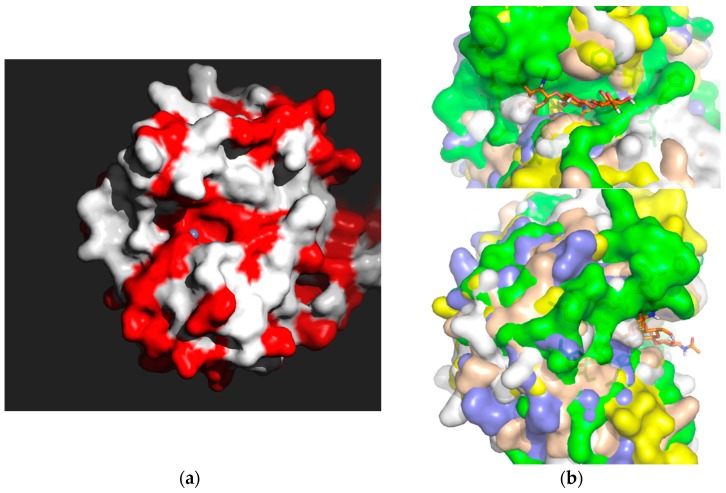
Figure 7.
Conservation of NodB binding sites. (a). Surface representation of the NodB PDA binding domain facing the active site with the Group 1 amino acid residue identity (in red) for Ba0331, Ba0330 and Bc0361. The metal (Zn) ion position in the active site is indicated with a grey sphere. (b). Surface representation of the NodB PDA binding site from selective B. anthracis and B. cereus structurally determined NodB domains superimposed (Bc1974 (yellow), Ba0331 (blue), Ba0330 (green), Bc1960 (beige), Ba0424 (white)) in two orthogonal views to highlight the similarities (shape) and detailed differences between the PDAs. A trisaccharide GlcNAc is modelled in Bc1974 (in orange sticks) [35]. The formation of the binding crevice (running horizontally) is shown as well as the detailed differences between the PDAs on the surface.

