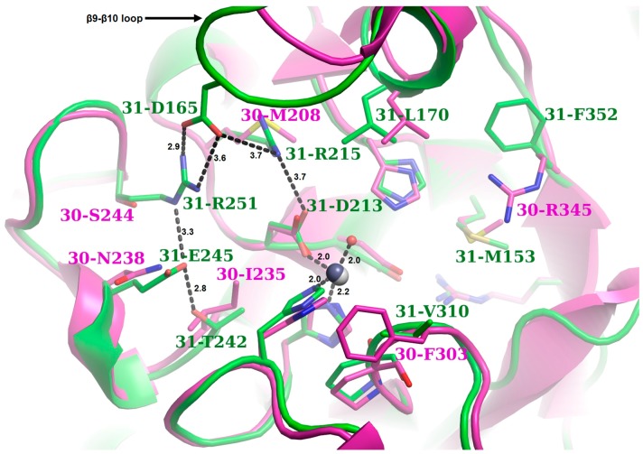
Figure 9.
Superposition of the NodB domain containing the binding sites of Ba0331 and Ba0330. Ba0331 is shown in green and Ba0330 is shown in magenta (acquired from PDBIDs: 4V33, 6GO1). The figure is centred on the zinc ion and therefore only the binding site grooves are shown. Τhe residues that either belong directly to an MT1-5 domain or lie within a 5 Å distance from one of these motifs are represented as sticks. The acetate ions were removed for clarity reasons. A black sphere represents the zinc ion in the active site for Ba0331 and a grey sphere shows the zinc ion for Ba0330. The zinc coordination sphere or the interacting network unique in Ba0331 is shown in black dashed lines (distances in angstroms). Only the different residues between Ba0331 and Ba0330 are labelled. A strong interacting network (on the left side of the figure), which corresponds to the upper rim of the binding groove, was observed in Ba0331. In contrast to the Ba0330/Bc0361 deacetylases where the particular side is occupied by hydrophobic or small amino acids, in Ba0331, a number of charged or hydrophilic residues were located. Strong interactions of D165 with positively charged residues stabilize the β9–β10 loop to a closed conformation, thus reducing the available volume of the binding groove. On the right side of the figure, which corresponds to the lower side of the binding groove, the presence of M153, L170 and F352 characterized the boundaries as hydrophobic.

