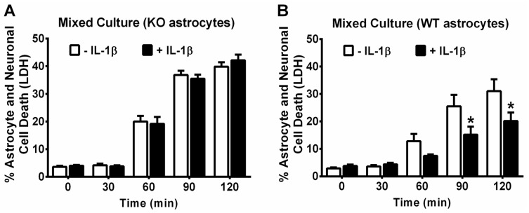
Figure 5.
IL-1β-mediated protection against t-BOOH-toxicity in mixed cultures is dependent on astrocyte activation. Wild-type neurons plated on astrocytes cultured from (A) il1r1−/− or (B) il1r1+/+ mice were treated with its vehicle (−IL-1β) or 10 ng/mL IL-1β (+IL-1β) for 48 h followed by treatment with 1.5 mM t-BOOH. Supernatants were collected for measurement of lactate dehydrogenase LDH at each time point, as indicated. Cultures were then washed free of t-BOOH, and medium collected again at 165 min (total time of exposure plus post-wash incubation) from each well. Values were summed and normalized to LDH released from cultures treated with 1.5 mM t-BOOH for 20–24 h (=100% cell death). Data are mean % cell death + SEM (A) There were no statistically significant group effects as determined by two-way ANOVA (n = 16 from 4 separate dissections); (B) p values were equal to 0.0035 for the IL-1β treatment effect, <0.0001 for the effect of time of t-BOOH exposure, and 0.0149 for the treatment × time interaction. An asterisk (*) depicts significant between-group difference (p < 0.05). Two-way ANOVA followed by Bonferroni’s test for multiple comparisons (n = 16 from 4 separate dissections). WT: il1r1+/+; KO: il1r1−/−.

