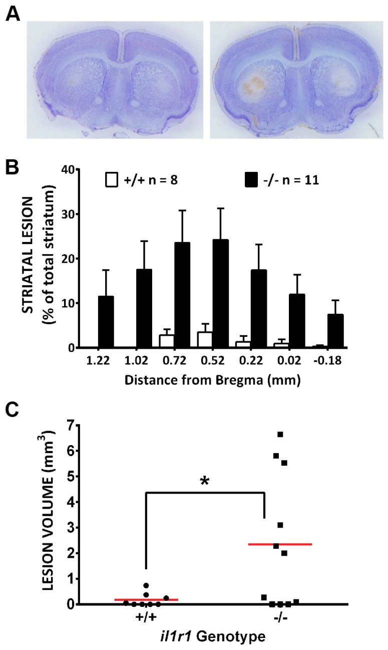
Figure 7.
Mice deficient in IL-1R1 signaling (il1r1−/−) have larger striatal lesions following systemic 3-NP treatment as compared to their wild-type littermate (il1r1+/+) controls. Male il1r1−/− (n = 11) and il1r1+/+ (n = 8) were injected twice daily with 3-NP as described in the methods. (A) Representative photomicrographs of thionin-stained sections (+0.52 from bregma) taken from 3-NP-treated il1r1+/+ (left) and il1r1−/− mice (right); (B) Comparison of lesion area between il1r1+/+ and il1r1−/− mice expressed as a percentage of total striatal area. A significant difference between genotypes occurs at every level of bregma as determined by repeated measures two-way ANOVA (p = 0.0163), followed by Sidaks post-hoc t-tests for multiple comparisons (p < 0.0001 at all levels); (C) Comparison of lesion volume between il1r1+/+ and il1r1−/− mice determined using the Cavalieri’s method. Data points represent the lesion volume (mm3) for each individual mouse, while a horizontal line represents the mean mm3 for each group. A one-tailed t-test with Welch’s correction revealed a significant difference (*) between genotypes at p = 0.01.

