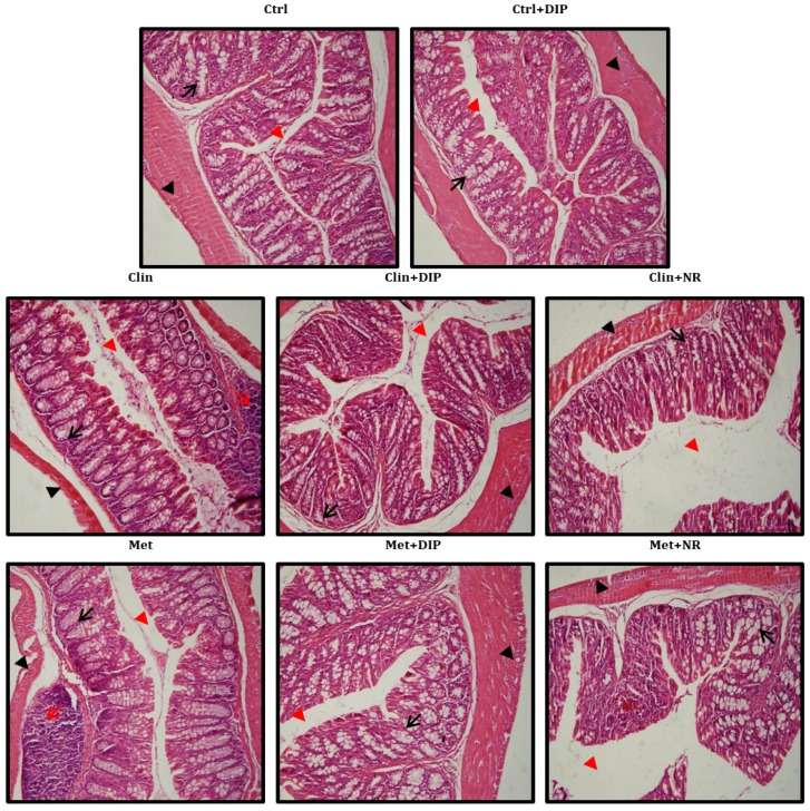
Figure 6.
Effect of DIP on colonic histological alterations in antibiotic-induced dysbiosis mice. Photomicrographs of hematoxylin and eosin (H&E)-stained distal colon sections. Shown are inflammatory cells (indicated with red arrows), the mucosal space (red arrow head), goblet and epithelial cells (black arrows), and the epithelium surface (black arrow head). Original magnification ×20, Scale bar: 100 µm.

