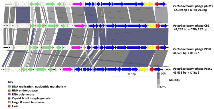
Figure 3.
Comparison of the genomes of the phages that form the proposed genus of ‘Phimunavirus’. Pectobacterium phage CB5 and Pectobacterium phages φM1, Peat1, and PP90 are shown using currently available annotations from Genbank, employing BLASTN and visualization with Easyfig. The genome maps display arrows indicating locations and orientation of ORFs among different phage genomes. Arrows have been color-coded describing their predicted roles (see key), and shading between the genome maps indicates the level of identity. Phage DTRs of unknown length marked with “?”.

