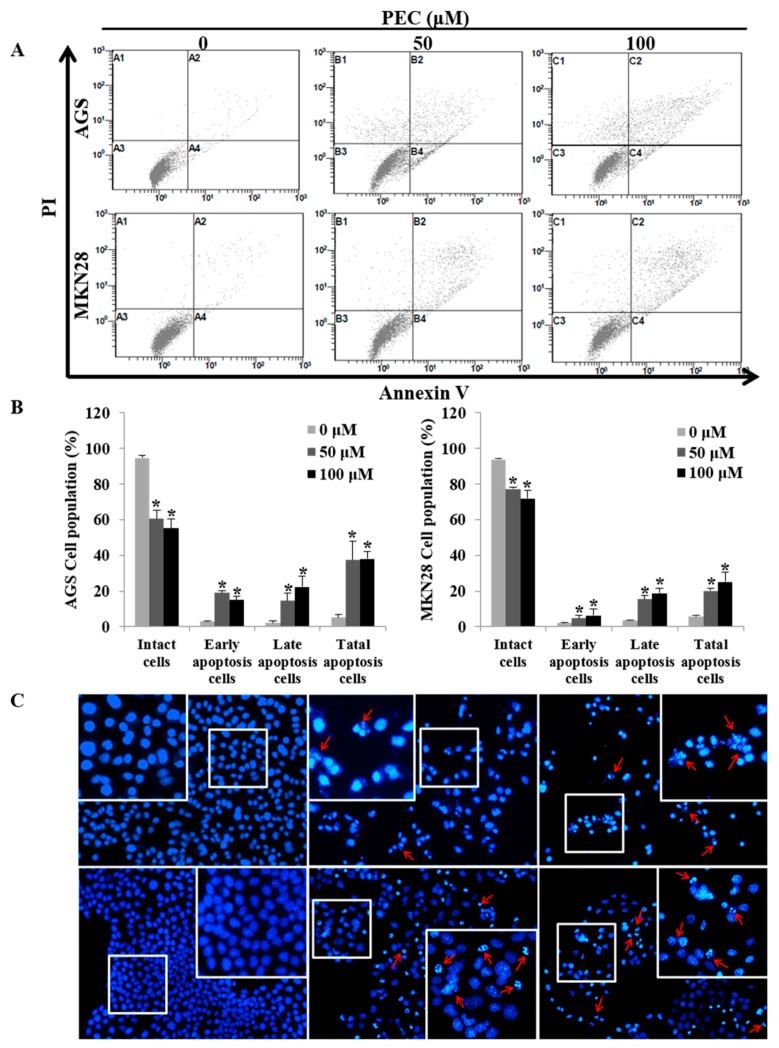
Figure 3.
PEC induces dose-dependent apoptosis in AGS and MKN28 cells. (A,B) Cells were treated with indicated concentrations of PEC for 24 h. Apoptotic cells were stained with Annexin V/PI kit and determined by flow cytometry. The cells were analyzed using CXP Software. Results are expressed as the mean ± standard deviation (SD) of at least three independent experiments. Statistical differences were analyzed with Student’s t-test (* p < 0.05 vs. control). (C) Both AGS and MKN28 cells were stained with Hoechst 33342 and examined under fluorescence microscopy (×400). (Red arrows showing bright blue regions indicate fragmented or condensed nuclei).

