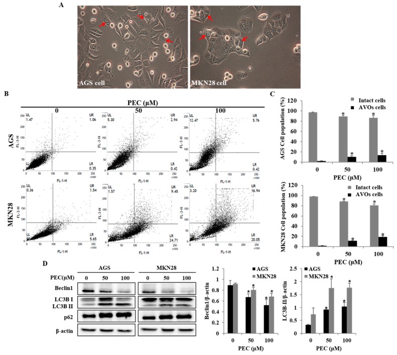
Figure 5.
PEC induces autophagy cell death in AGS and MKN28 cells. (A) Morphology of both AGS and MKN28 cells was examined under light microscopy (×400) after treated with PEC (100 μM). (The red arrows indicate the autophagic vacuoles.) (B) The cells were treated with PEC at 0, 50 and 100 μM concentrations for 24 h. After incubation, the cells were stained with 5 μg/mL acridine orange for 15 min and collected in phenol red-free growth medium. Green (510 ± 530 nm) and red (650 nm) fluorescence emission illuminated with blue (488 nm) excitation light were measured with a flow cytometer. PEC induced dose-dependent AVO formation in AGS and MKN28 cells. (UR and UL: AVOs cells; LL and LR: Intact cells). (C) AGS and MKN28 were treated with indicated concentrations of PEC or 24 h. The cell lysates were subjected to SDS-PAGE and analyzed by immune-blotting. Densitometry analyses of Beclin1, LC3, and p62 protein expressions are presented. Results are expressed as the mean ± standard deviation (SD) of at least three independent experiments. Statistical differences were analyzed with Student’s t-test (* p < 0.05 vs. control).

