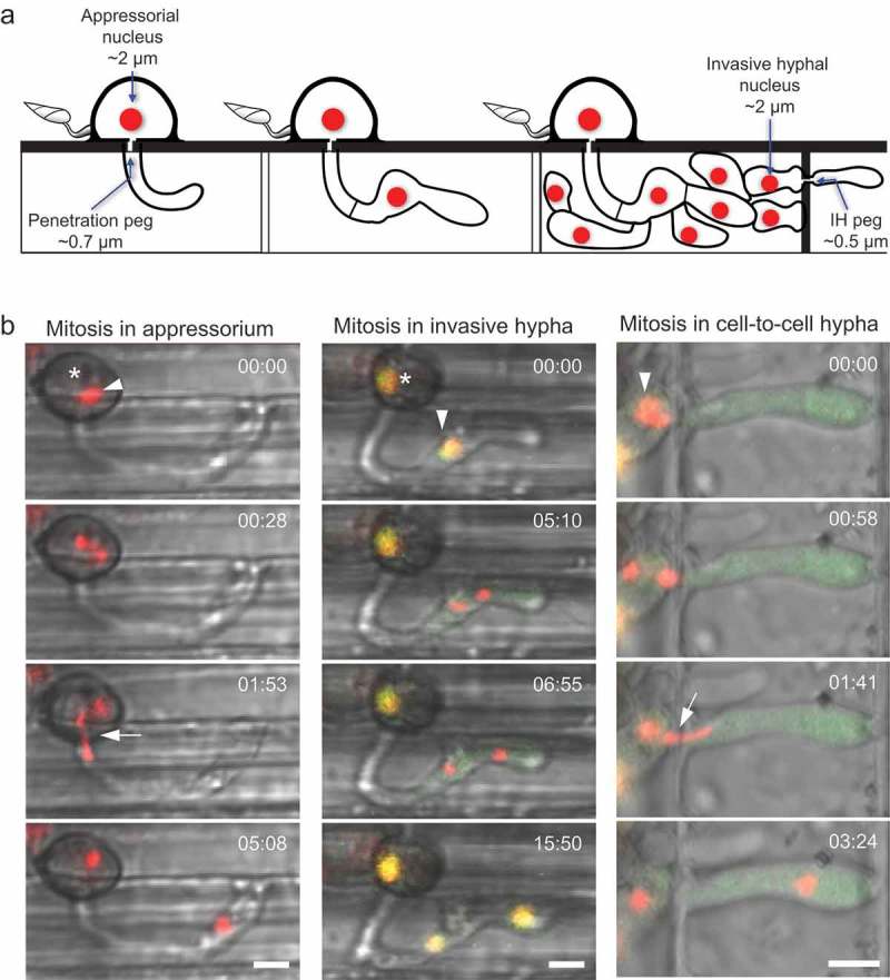
Figure 2.

Mitotic migration of M. oryzae nuclei during early rice blast infection. (a) Schematic diagram summarizing key cellular structures and mononuclear positioning during plant invasion. The nucleus in the appressorium (interphase diameter of ~2 μm) must traverse the constricted penetration peg (diameter of ~0.7 μm) for final receipt in the incipient primary hypha. Once inside the first-invaded cell, the primary hypha becomes bulbous to form invasive hyphae (IH). To move into adjacent rice cells, IH seek out pit fields and develop a constricted IH peg (diameter of ~0.5 μm). (b) A time-lapse series of nuclear dynamics at three distinct stages of early rice blast infection. Asterisks denote the appressorium, arrowheads label a nucleus about to undergo mitotic nuclear migration, and arrows highlight extreme nuclear morphology during confined nuclear migration through peg structures. (Left: merge of bright-field and H1-tdTomato.) Mitosis begins in the appressorium, and the daughter nucleus becomes highly constricted and elongated during confined mitotic nuclear migration through the penetration peg. The original nucleus remains located in the appressorium throughout this event. GFP-NLS dynamics (data not shown) confirms nuclear migration occurs during intermediate mitosis at this infection stage (Jenkinson et al. 2017). (Middle panel: merge of bright-field, GFP-NLS, and H1-tdTomato.) During mitosis in the invasive hypha, the interphase nucleus appears yellow due to colocalisation of H1-tdTomato and GFP-NLS in the nucleus. After onset of mitosis, GFP-NLS disperses into the cytoplasm, and the nucleus undergoes an unconfined nuclear migration. Following receipt of the nucleus into the new invasive hypha cell, mitosis ends, and GFP-NLS is fully reimported back into the nucleus. (Right panel: merge of bright-field, GFP-NLS, and H1-tdTomato.) Here, confined nuclear migration through the IH peg occurs. In early mitosis, the sister chromatids separate and during presumed anaphase B, a single daughter nucleus undergoes confined nuclear migration through the constricted IH peg. The daughter nucleus again becomes spherical and continues to migrate to the tip of the IH in the second-invaded cell prior to GFP-NLS reimport into the nucleus. Times are shown in minutes:seconds. Bars = 5 μm. This figure is modified from Jones et al. (2016a) and Jenkinson et al. (2017).
