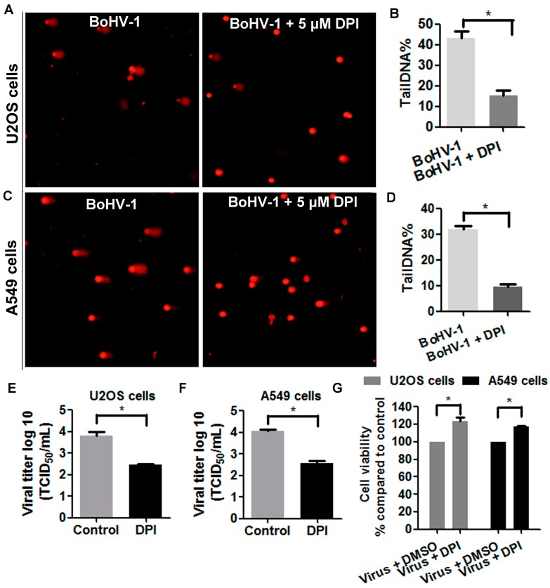
Figure 6.
ROS was involved in BoHV-1-induced DNA damage in human tumor cells. (A,C) Both U2OS cells (A) and A549 cells (C) were infected with BoHV-1 (MOI = 0.1) and treated with DPI (5 μM) or DMSO control. At 48 h after infection, DNA damage in individual cells was detected with comet assay. The images shown represent three independent experiments (Magnification ×200). (B,D) Three hundred cells from both U2OS cells (B) and A549 cells (D) treated with or without DPI were randomly selected for the analysis of tailDNA% with software CASP. Results are means of three independent experiments. *, Significant differences (P < 0.05) in tailDNA%, as determined by a Student t test. (E,F) Both U2OS cells (E) and A549 cells (F) were infected with BoHV-1 (MOI = 0.1) and treated with or without DPI (5 μM). At 48 h after infection, the virus titers were detected using MDBK cells. *, significant differences (P < 0.05), as determined by a Student t test. (G) Both U2OS cells and A549 cells were infected with BoHV-1 (MOI = 0.1) and treated with DPI (5 μM) or DMSO control. At 48 h after infection, the cell viability was determined by Trypan-blue exclusion test. Results are means of three independent experiments. *, Significant differences (P < 0.05), as determined by a Student t test.

