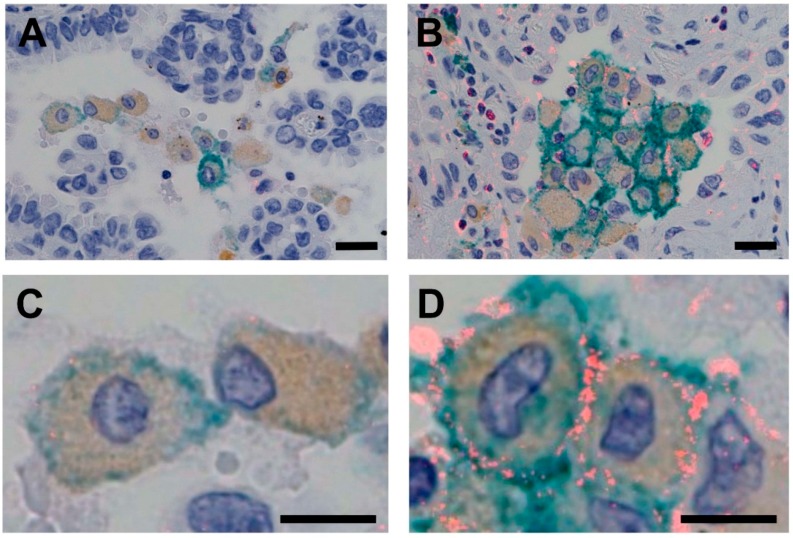
Figure 1.
Triple-stained images for PID, DAB and HistoGreen. CSF1R-expressing TAMs stained positive for CSF1R (red), CD68 (brown) and CD163 (green). (1) TAMs with low expression levels of CSF1R (A: low magnification, scale bar = 20 µm; C: high magnification, scale bar = 10 µm). (2) TAMs with high expression levels of CSF1R (B: low magnification, scale bar = 20 µm; D: high magnification, scale bar = 10 µm). DAB, diaminobenzidine; PID, phosphor-integrated dot; TAM, tumor-associated macrophage.

