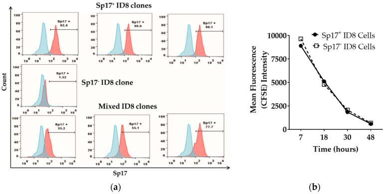
Figure 1.
Sperm protein 17 (Sp17) is expressed in a subset of ID8 cells in vitro. (a) Sp17 expression in selected ID8 sub-clones: Sp17+ (top panel, three sub-clones obtained), Sp17− (middle panel, one sub-clone obtained), and mixed clones (bottom panel, three representative sub-clones), analyzed by flow cytometry. Data is shown as histogram of Sp17 expression (red) over isotype control (blue) for each of the ID8 sub-clone presented here. The X-axis shows the fluorescence intensity and the Y-axis shows count. All cells were stained intracellularly with an anti-Sp17 antibody; (b) in-vitro growth of the Sp17+ and Sp17− ID8 cells. The Sp17+ and Sp17− cloned ID8 cells were stained by carboxyfluorescein succinimidyl ester (CFSE) and cultured for 7, 18, 30, and 48 h. CFSE fluorescence was assessed by flow cytometry at each time point.

