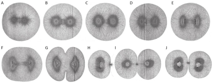
Figure 3.
First cleavage in the early embryo of Echinus microtuberculatus. Sea urchin embryos were fixed in picric-acetic acid and stained with iron haematoxylin. (A) Chromosomes are arranged at the metaphase plate (equatorial plane). Centrosomes are spherical. (B) Centrosomes increase in size while homologs become separated. (C) Centrosomes start to flatten in the direction parallel to the division axis. (D) Flattened centrosomes change to discoidal form. (E) Centrosomes adopt a discoidal shape. (F) Spindle and egg elongate. Chromosomes are visible and centrosomes start to divide. (G) Cytokinesis of the embryo starts. (H,I) Embryonic daughter cells are formed (side-view (H) and cross-section/top-view (I) according to division axis). (J) Each daughter cell contains a nucleus and two centrosomes. Reproduced from [2].

