Figure 1.
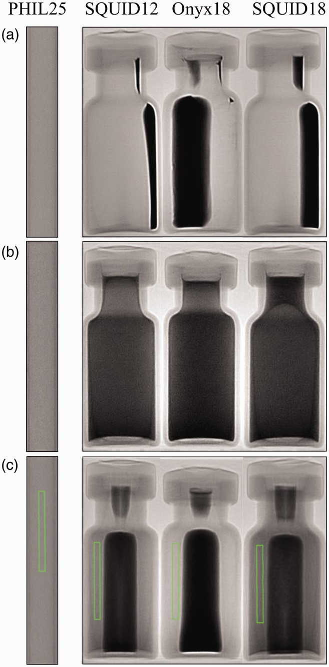
(a) Onyx 18®, SQUID 12®, SQUID 18®, and PHIL 25® prior to preparation. Tantalum sediment is clearly visible in the vials of Onyx 18®, SQUID 12®, and SQUID 18®. (b) Studied embolic agents one minute after preparation: Tantalum is uniformly distributed. (c) After 30 minutes, tantalum can be seen settling within the middle of each vial. Additionally, regions of interest (ROIs) used to measure pixel data seen are placed within each liquid embolic. These ROIs were copied into the exact same position for all measurements.

