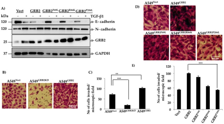
Figure 4.
Functional N-terminal and C-terminal SH3 domains are critical for reducing E-cadherin expression and promoting invasion of A549 cells, respectively. (A) A549 cells were infected with lentivirus particles generated using vector, or plasmids expressing WT GRB2 or its mutants (GRB2P49L, GRB2R86K and GRB2P206L), selected with puromycin, and EMT was as described in Figure 1. Cells were then lysed and the protein extracts were subjected to Western blot analysis by probing with antibodies against E-cadherin, N-cadherin, and GRB2. GAPDH was used as the loading control; (B) Matrigel-coated invasion assay inserts were seeded with 2 × 10⁵ cells of A549Vect, A549GRB2KD and A549GRB2 in 200 µL of serum-free media on the upper chamber of the insert. The well below was filled with complete media containing 20% FBS. The cells were incubated for 40 h at 37 °C, the cells which had invaded through the Matrigel were fixed, washed and stained with crystal violet, and imaged under a 10× objective lens; (C) Cells in four random fields per sample were counted to quantify the number of cells that had invaded. Three independent sets of experiments were carried out to calculate significance. (** p < 0.01, *** p < 0.001); (D) Matrigel-coated invasion assay inserts were seeded with 2 × 10⁵ cells of A549Vect, A549GRB2, A549GRB2P49L, A549GRB2R86K and A549GRB2P206L per 200 µL of serum-free media in the upper chamber of the insert. The well below was filled with complete media containing 20% FBS. The cells were incubated for 40 h at 37 °C; the cells that had invaded through the Matrigel were fixed, washed and stained with crystal violet and imaged under a 10× objective lens; (E) Four random fields per sample were imaged and the cells were counted to quantify the number of cells that had invaded through the Matrigel. Three independent sets of experiments were carried out to calculate significance. (** p < 0.01, *** p < 0.001).

