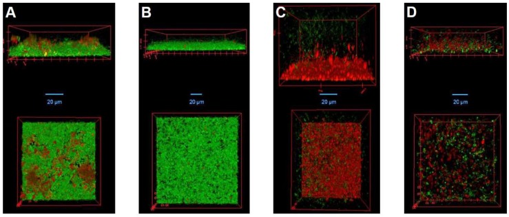
Figure 5.
Confocal laser scanning micrographs of S. aureus biofilms treated with DA7, LysK, or a combination of both agents. SA113 biofilms were grown in IBIDI µ-slides for 20 h at 19 °C and then treated for 2 h with: buffer (A); 0.625 nM DA7 (B); 1.25 µM LysK (C); or a combination of 0.156 nM DA7 and 0.938 µM LysK (D). Residual biofilms after treatment were stained with LIVE/DEAD stain and then visualized by CLSM. Side views (top); and top views (bottom) of biofilm 3D reconstructions are shown. Live cells are depicted in green and dead cells as well as extracellular DNA in red.

