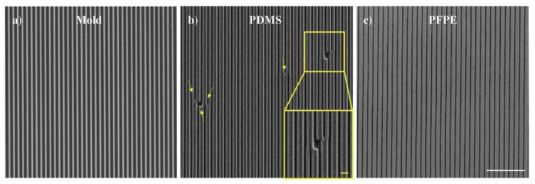
Figure 1.
Scanning electron microscope representative images of (a) the 600 nm-periodic nano-grating initial mold, (b) the PDMS replica, and (c) the PFPE replica. Scale bar: 5 μm. Yellow arrows highlight the presence of defects in the PDMS replica. Inset of (b) zoomed image of a representative area with defects, scale bar = 1 μm.

