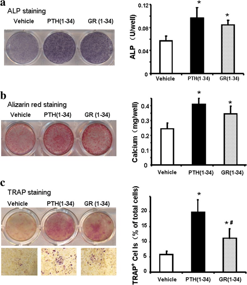
Fig. 6.

The effects of hPTH(1–34) and GR(1–34) on osteogenesis and osteoclastogenesis in bone marrow cells of ORX mice. At the 2nd week of administration, ALP activity of the cells in osteogenic medium were stained and measured with a BCIP/NBT Color Development Kit (a); the mineralized nodules were stained with Alizarin Red S solution and calcium deposition was quantified spectrophotometrically (b). At the 1st week of administration, TRAP staining was performed and the percentage of TRAP+ cells to total cells were counted (c). [Three independent experiments were repeated for (a & b) and six for (c); 10 fields at 100 magnitude were randomly selected for TRAP+ cell counting; *P < 0.05 for hPTH(1–34) &GR(1–34) vs. vehicle; #P < 0.05 for hPTH(1–34) vs. GR(1–34)]
