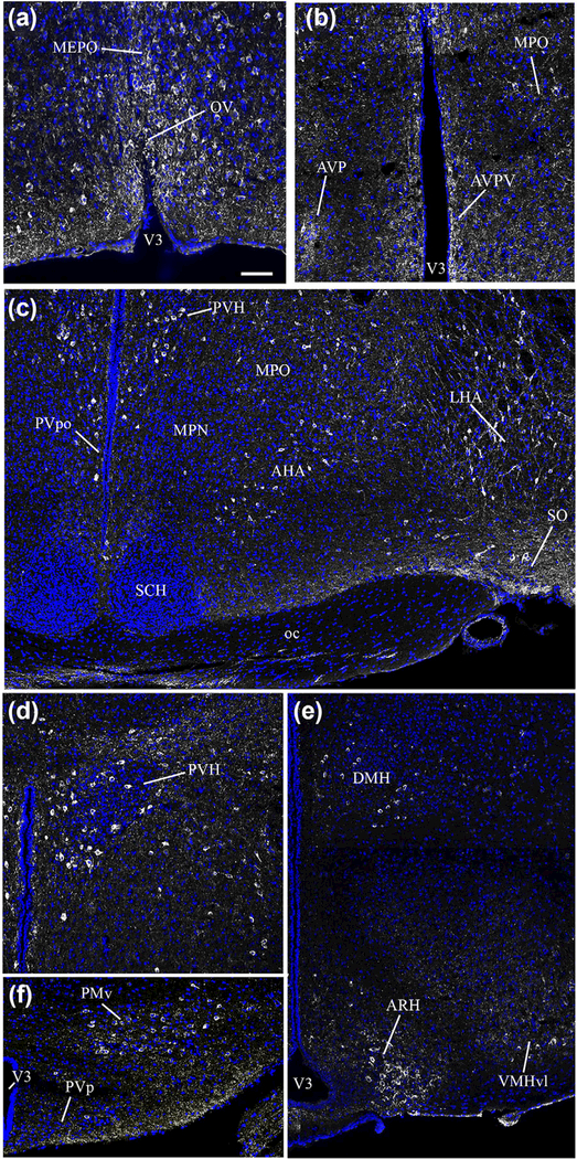
FIGURE 1.
Photomicrographs showing the distribution of nNOS immunoreactivity in distinct hypothalamic nuclei as seen in coronal sections of the adult female mouse brain. nNOS immunoreactivity (white labeling) is readily visualized in neurons of the preoptic area; labeling is evident in the regions of the organum vasculosum laminae terminalis (OV, a), median preoptic nucleus (MEPO, a), anteroventral preoptic nucleus (AVP, b), anteroventral periventricular nucleus (AVPV, b), and medial preoptic region (MPO, b). nNOS immunoreactivity was also detected in the anterior hypothalamic nucleus (AHN, c), lateral hypothalamic area (LHA, c) and supraoptic nucleus (SO, c), with rare neurons also present in the periventricular nucleus of the hypothalamus (PVp, f). nNOS-expressing neurons were also visualized in the paraventricular nucleus of the hypothalamus (PVH, d), the dorsomedial nucleus of the hypothalamus (DMH, e), the ventrolateral part of the ventromedial nucleus of the hypothalamus (VMH, e), the arcuate nucleus of the hypothalamus (ARH, e) and the ventral premammillary nucleus (PMv, f). Sections are counterstained using Hoechst (blue) to visualize cell nuclei and identify the morphological limits of each hypothalamic structure. 3V: third ventricle. Scale bar =100 μm in (a), (b), (d) and (f), and 200 μm in (c) and (e)

