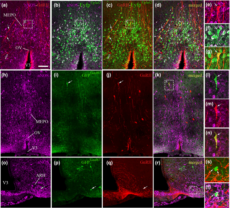
FIGURE 6.
Representative images showing nNOS and GnRH immunoreactive neurons in EYFPVgut2 and GFPGad67 female mice. Colocalization of the enhanced yellow fluorescent protein (YFP-IR, green) is evident in both nNOS-immunoreactive cells (purple; b, f) and GnRH-immunoreactive neurons (red; c, g) in the preoptic region of adult female EYFPVgut2 mice (a-g). Expression of green fluorescent protein (GFP) driven by the Gad67 promoter is absent in nNOS-immunoreactive cells (purple) of the preoptic region (h-i), while it is occasionally expressed in GnRH-immunoreactive neurons (red; i-n). In the arcuate nucleus of the adult female hypothalamus (ARH), nNOS-immunoreactive neurons expressing GFPGad67 (o-p, t) are surrounded by GnRH-immunoreactive fibers (red; q, s, t). Scale bar = 100 im (25 im in insets)

