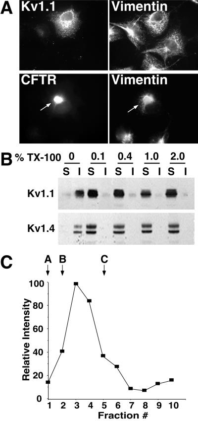
Figure 2.
Folding and assembly of Kv1 channels. (A) COS-1 cells expressing Kv1.1 (Upper) or CFTRΔF508-GFP (Lower) were fixed, permeabilized, and stained with anti-Kv1.1 (Upper Left) and anti-vimentin (Upper and Lower, Right). Arrows show aggresome formation, indicated by vimentin collapse into ring-like structures. (B) COS-1 cells expressing Kv1.1 (Upper) or Kv1.4 (Lower) were harvested and permeabilized in lysis buffer containing 0%, 0.1%, 0.4%, 1.0%, or 2.0% TX-100 detergent. Soluble (S) and insoluble (I) fractions were separated by centrifugation and analyzed by SDS/PAGE and immunoblotting. (C) COS-1 cells expressing Kv1.1 were harvested and permeabilized with 1.0% TX-100. Soluble lysates were fractionated on a 5–50% linear nondenaturing sucrose gradient. Fractions were subjected to immunoprecipitation, SDS/PAGE, immunoblotting, and densitometry. Gradient controls are as follows: A, carbonic anhydrase; B, BSA and alcohol dehydrogenase; and C, apoferritin.

