. 2018 Mar 31;31(1):23–27.

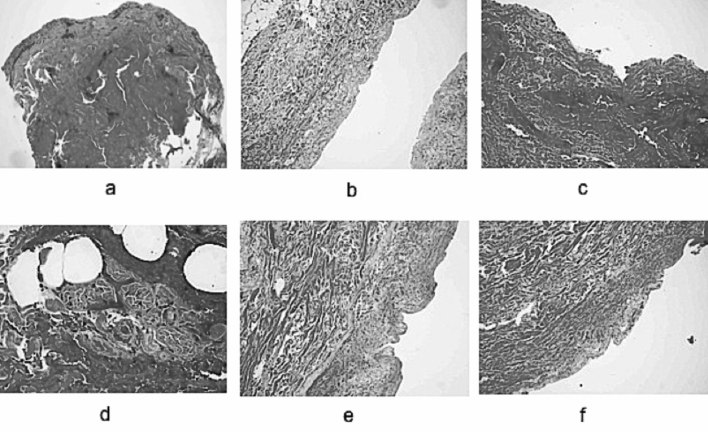
Fig. 3. Biopsy sections before and after debridement with NexoBrid® on partial thickness burn wounds. a) biopsy before NexoBrid. De-epithelialized dermis with areas of coagulative necrosis; b) biopsy after NexoBrid. The dermis is de-epithelialized and the most superficial area looks homogenized; c) post-debridement biopsy on ecchymotic area. The superficial dermis has become denser and infiltrated with erythrocytes; d) at 40x magnification, capillary disruption with blood extravasation is observed. Inflammatory infiltrates and partially damaged annex structures; e) homogenized superficial dermis; deep dermis with normal structure; f) the most superficial dermis shows the presence of areas with residual thermal necrosis and partially viable skin annexes.

Fig. 3