
Figure 1.
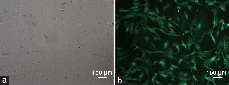
Morphology of cultured ADSC- and GFP-positive cells. (a) Passage 3 ADSCs were in fibroblast-like shape. (b) Fluorescence images of passage 3-infected ADSCs at an MOI of 100. Scale bars = 100 μm. ADSC: adipose-derived stem cell; MOI: multiplicity of infection; GFP: green-fluorescent protein.