
Figure 6.
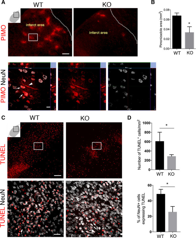
RGS5 (regulator of G-protein signaling 5) loss in pericytes is associated with reduced hypoxia and increased neuronal survival after stroke. A, Representative confocal images showing pimonidazole (PIMO) staining in the infarct area of wild type (WT) and knockout (KO) mice; scale, 50 μm. Arrows indicate NeuN expressing neurons positive for PIMO in the framed area in (A), scale bar, 10 μm. B, Quantification of PIMO-positive area in the infarct area of WT and KO mice; mean±SD (n=3, *P<0.05, Student t test). C, Confocal images showing TUNEL+ (terminal deoxynucleotidyl transferase dUTP nick end labeling) cells (top; scale bar, 50 μm) and TUNEL+ neurons (NeuN, bottom; scale bar, 20 μm) in WT and KO mice. D, Quantification shows significantly less TUNEL+ cells in KO vs WT mice; mean±SD (n=3, *P<0.05 Student t test). The percentage of NeuN cells colabeling with TUNEL is significantly lower in KO than WT mice; mean±SD (n=3, *P<0.05, Student t test).