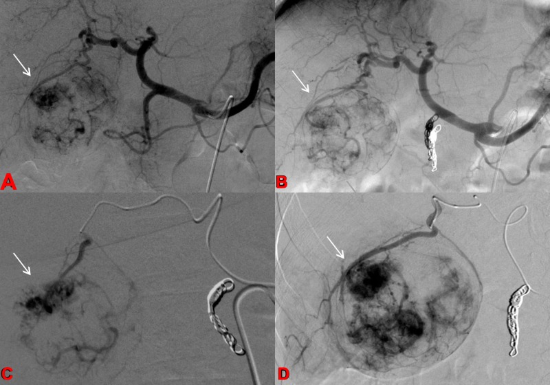
Figure 2. Pretreatment Angiogram.
Celiac arteriograms before and after embolization of the gastroduodenal artery (a-b) demonstrate conventional celiac vascular anatomy with tumor blush in the right hepatic lobe corresponding to the known hepatocellular carcinoma (HCC). C-D: Selective arteriograms of the hepatic arterial branches supplying the hypervascular HCC.

