Figure 1.
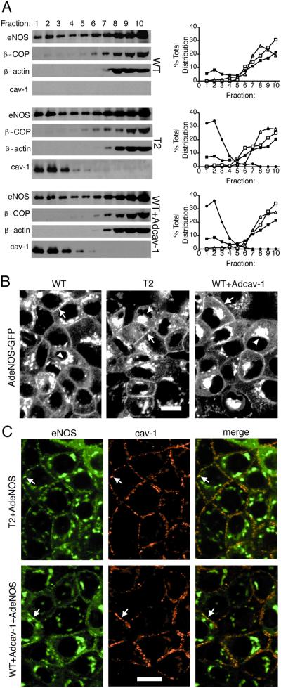
Ectopic expression of cav-1 in FRT cells results in caveolae assembly but has no effect on eNOS trafficking. (A) Cav-1-negative parental (WT) FRT cells, T2, or parental FRT cells infected with Adcav-1 (WT + Adcav-1) were infected with AdeNOS, and isolation of raft domains was performed on bottom-loaded sucrose gradients as described. The relative enrichments in eNOS, β-COP, β-actin, and caveolin in equal volumes of each fraction were assessed. Right is quantitative densitometry reflecting the distribution of eNOS (filled box), β-COP (open box), β-actin (open triangle), or cav-1 (filled circle) throughout the gradient. (B) WT, T2, or parental FRT + Adcav-1 were infected with AdeNOS-GFP, and the localization of eNOS-GFP was examined in living cells. Arrows depict eNOS-GFP in the plasma membrane, and arrowheads show eNOS-GFP in perinuclear structures. (C) The localization of eNOS (Left) and cav-1 (Center) were performed by immunofluorescence microscopy in T2 and FRT cells infected with AdeNOS virus. Far Right shows the merged images. Arrows denote examples of colocalization of eNOS and cav-1 in plasma membrane. Calibration bars = 20 μm for B and C. Please refer to Table 1 for morphometric evaluation of caveolae and other vesicle populations.

