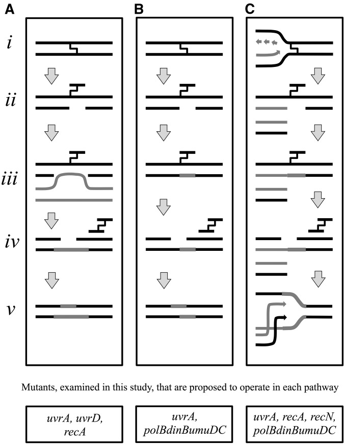
Figure 1.
Predominant models for the repair of DNA interstrand cross-links in the literature. (A) Nucleotide excision repair and homologous recombination. The interstrand cross-link (i) is initially incised by the nucleotide excision repair complex (ii), before the gap on the incised strand is filled in by recombination using a sister chromosome (iii). Nucleotide excision repair could then in theory make a second round of incisions on the opposing strand (iv), which could be filled in using the “newly formed” complementary strand as a template (v) (adapted from Cole 1973a; Van Houten et al. 1986; Sladek et al. 1989b). (B) Nucleotide excision repair and translesion synthesis. The interstrand cross-link (i) is initially incised by the nucleotide excision repair complex (ii) before translesion synthesis fills in the gap (iii). Nucleotide excision repair then makes a second round of incisions to remove the lesion (iv) before the remaining gap is resynthesized using the newly formed complementary strand as a template (v) (adapted from Berardini et al. 1997). (C) Excision-mediated double-strand break repair. Following an encounter with a replication fork (light arrows) (i), incision of the interstrand cross-link by nucleotide excision repair results in a double-strand break (ii). Translesion polymerases restore the incised template (iii) before a second round of nucleotide excision repair removes the cross-link and restores the template (iv). Recombination then repairs the double-strand break to restore the replication fork indicated by arrows (v) (adapted from De Silva et al. 2000; Bessho 2003; Niedernhofer et al. 2004). Genes examined in this work that are proposed to operate in each pathway are listed below each model.

