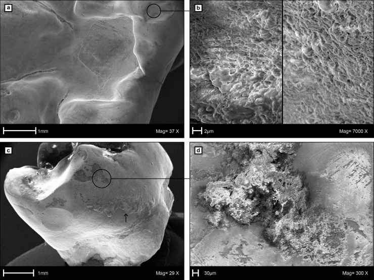
Fig 6. Scanning Electron Microscopy (SEM) images of the caries cavity and calculus on the left m1 of LMK-Pal 5508.
a, Occlusal view into the caries cavity. b, Surface details of occlusal dental calculus close to the caries cavity. c, Supra-gingival calculus (above arrow) on the mesiobuccal enamel surface. d, Surface details of mesiobuccal calculus and scratch dominated microwear on the enamel surface.

