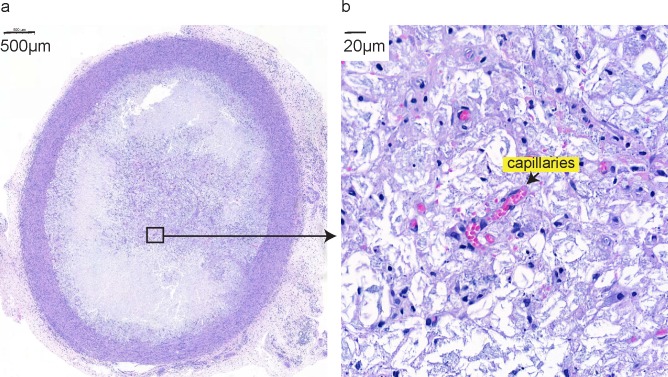
Fig 3. Hydrogel monitoring.
Hydrogel deposits explanted for analyses on POD 9 show fibrotic capsule formation (a) and vascularization–capillaries formed inside the hydrogel deposit, indicated by an arrow (b). Shown are representative histological hematoxylin and eosin stained sections of hydrogel deposits at 3x (a) and 50x (b) magnification.

