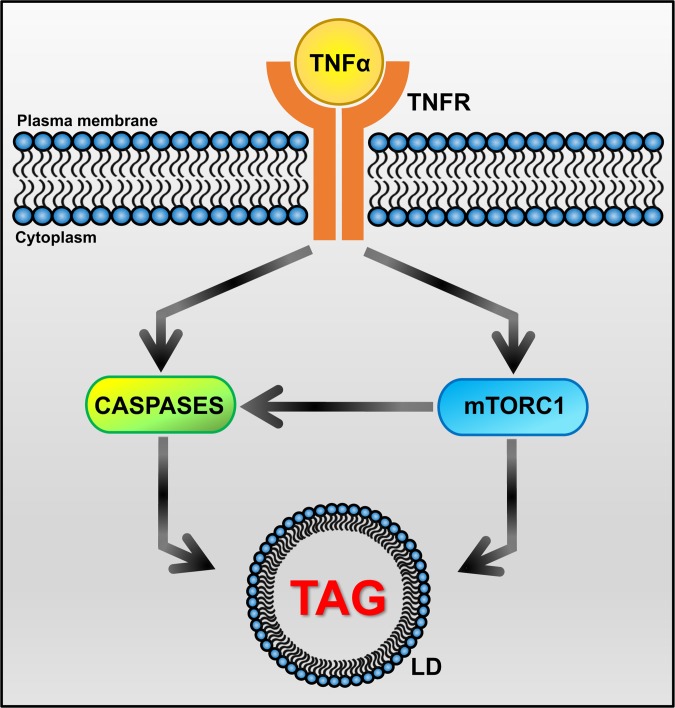
Fig 7. Key nodes of the signaling network involved in TAG accumulation in tuberculous macrophages.
Infection of macrophages with M. tuberculosis induces production of TNFα, which has both autocrine and paracrine effects. TNFα binding to its surface receptors (TNFR1 and TNFR2) triggers the activation of intracellular caspase and mTORC1 pathways. Both mTORC1 and caspases directly interact with autophagy effectors to inhibit autophagic flux [42, 52, 109]. So does M. tuberculosis infection [110]. In addition, mTORC1-dependent block of autophagy can also activate the caspase cascade through accumulation of p62, which binds and activates caspase 8 [111]. When autophagy is blocked, lipid droplets are not degraded and accumulate in the cytoplasm. Our observation that treatment with TNFR neutralizing antibodies relieves the inhibition of autophagy by M. tuberculosis infection (S6 Fig) further supports inhibition of autophagy as a driver of lipid droplet accumulation in tuberculous macrophages. In addition, caspase activation also leads to mitochondrial dysfunction [43], which results in accumulation of lipid droplets due to reduced fatty acid utilization [39]. Effects on TAG biosynthesis may also be involved, since mTORC1 induces expression of SREBF1 [112–114] and caspases activate SREBPs [40, 41]. Arrowhead: positive regulation. LD: lipid droplet.

