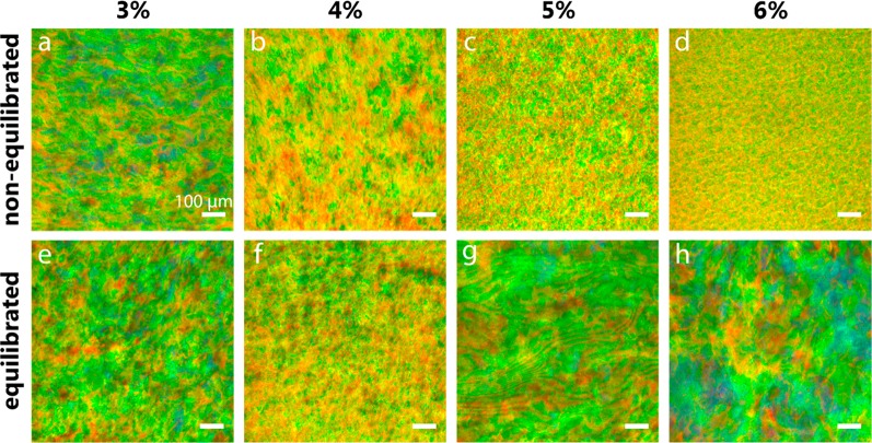
Figure 2.
Reflected-light microscopy images obtained with the light beam incident perpendicular to the film plane. (a–d) CNC films obtained directly after drying for 2 (d) to 4 days (a). (e, f) CNC films obtained after equilibrating the suspension for 7 days postcasting and prior to drying. The microscope images correspond to samples (a) N3, (b) N4, (c) N5, (d) N6, (e) E3, (f) E4, (g) E5, and (h) E6. The scale bars correspond to 100 μm.

