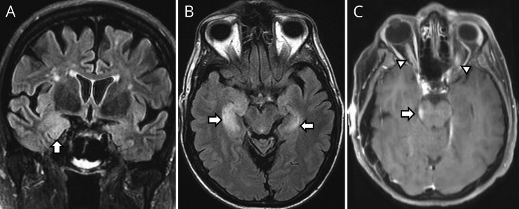
Figure. MRI of posttransplant autoimmune encephalitis.
(A) Right mesial temporal T2-hyperintensity (arrow) on coronal Fluid-attenuated inversion recovery (FLAIR) in a patient with posttransplant anti-NMDA receptor encephalitis. (B) Bilateral (right more than left) mesial temporal T2-hyperintensity (arrows) in a patient with posttransplant anti-AMPA receptor encephalitis. (C) Concurrent midbrain enhancement (arrow) and bilateral optic nerve enhancement (arrowheads) in patient with posttransplant myelin oligodendrocyte glycoprotein antibody disease.

