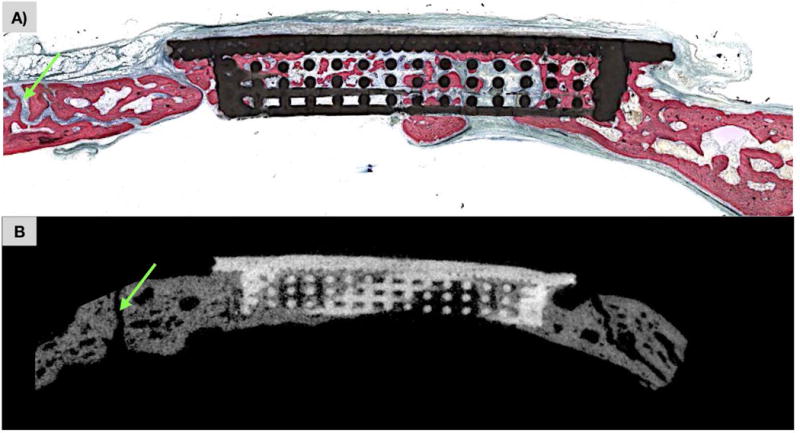
Figure 4.

a) Non-decalcified histologic section calvarial defect replaced by scaffold at t=8 weeks. New bone formation (pink) is observed through scaffold (black) porosity with patent cranial suture (green arrow). b) MicroCT slice corresponding to histologic slice, with scaffold construct (white) and new bone formation (grey) throughout scaffold interstices with patent cranial suture (green arrow).
