Fig. 1.

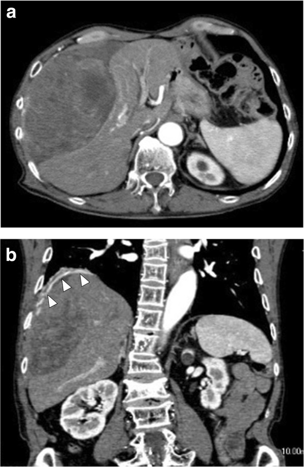
Computed tomography image showing a large subphrenic tumor. The heterogeneously enhanced tumor was well demarcated from the liver (a). The feeding artery originated from the diaphragm (arrowhead) (b)

Computed tomography image showing a large subphrenic tumor. The heterogeneously enhanced tumor was well demarcated from the liver (a). The feeding artery originated from the diaphragm (arrowhead) (b)