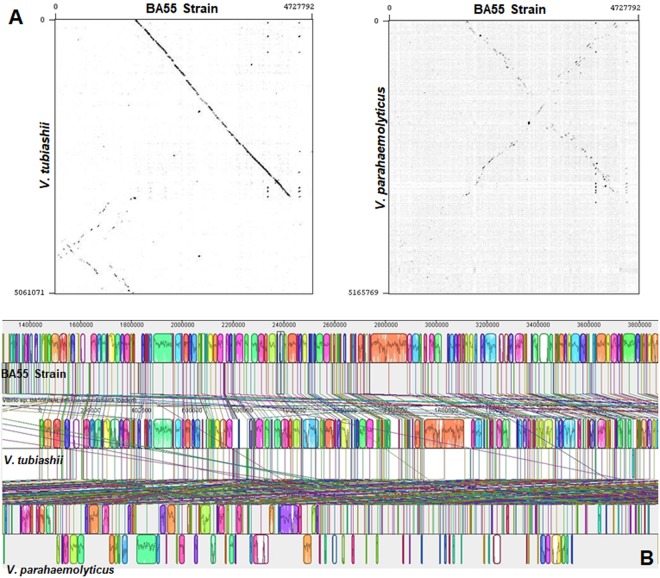
Figure 7.
Whole genome alignments between V. punensis strain BA55, V. tubiashii strain ATCC 19109 and V. parahaemolyticus RIMD 2210633. (A) Dot-plots of nucleotide identities of the BA55 strain against V. tubiashii strain ATCC 19109 (left) and BA55 strain against V. parahaemolyticus RIMD 2210633 (right). (B) Nucleotide-based multiple genome alignment of the same genomes. Homologous blocks are shown as identically colored regions and linked across the genomes. Rearrangements are shown as homologous regions in different genomic locations.

