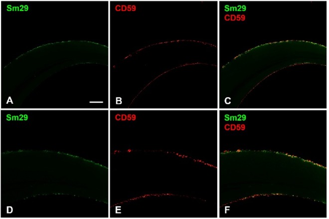
Figure 6.
Confocal images localizing Sm29 and CD59. Freshly perfused adult worms were fixed for whole mount assays with paraformaldehyde and incubated with primary antibodies and Alexa Fluor secondary antibodies before imaging on confocal microscope. (A,D) Anti-Sm29 antibodies confirmed the presence of Sm29 (in green) at the surface of the tegument as previously described13. (B,E) Anti-CD59 antibodies showed that CD59 (in red) was also found at the surface of the worms. (C,F) Merge of anti-Sm29 (in green) and anti-CD59 (in red) revealed that Sm29 and CD59 co-localized at the tegument surface of adult worms (in yellow).

