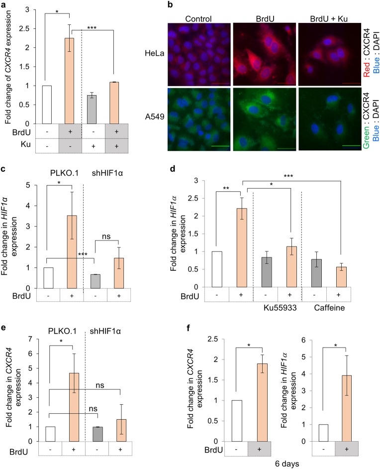
Fig. 2.
DNA damage response and CXCR4 expression. a Effect of ATM kinase inhibition on CXCR4 levels during DNA damage. CXCR4 expression analysis was performed by qRT-PCR in HeLa cells treated with BrdU and ATM kinase inhibitor as indicated. b Immunofluorescence analysis of CXCR4 expression. HeLa and A549 cells treated as indicated and probed for CXCR4 expression as described in Fig. 1E. Scale bar = 40 µm (n = 5). HeLa cells were probed with TRITC conjugated anti-CXCR4 antibody (red), while A549 cells with FITC–conjugated anti-CXCR4 antibody (green). c Analysis of HIF1α expression during DDR. Expression analysis of HIF1α in HeLa cells containing vector alone for HIF1α targeting shRNA after treatment with BrdU was performed by qRT-PCR (n = 3). d Effect of ATM kinase inhibition on HIF1α expression. HF-hTERT cells were treated with BrdU and treated with either Ku or caffeine. HIF1α expression was evaluated by qRT-PCR. (n = 3). e Analysis of CXCR4 expression wrt HIF1α levels. CXCR4 expression during BrdU induced DDR was analysed in HeLa cells stably expressing shRNA against HIF1α by RT-PCR (n = 3). f CXCR4 and HIF1α expression analysis after prolonged BrdU treatment. Expression analysis of CXCR4 (left) and HIF1α (right) was performed in HeLa cells treated with BrdU for 6 days by qRT-PCR and compared with untreated cells (n = 3). For all experiments results shown are mean ± s.e.m. p value ns < 0.5; *p ≤ 0.05; ***p ≤ 0.001 (Student’s t-test; n = 3)

