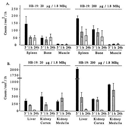
Figure 2.
Quantitative profile of radioactivity in the spleen, bone, and muscle (A) and liver, renal cortex, and renal medulla (B) of rats at 5 min (histograms 5′), 1 h, and 24 h after i.v. injection of 20 and 200 μg [3H]-labeled HB-19 (1.8 MBq or 50 μCi each). Direct quantification of sagittal whole-body sections was assessed by using the β-radio imager. The number of β-particles emitted per area was counted for 50 h and expressed as counts per mm2 per 1 h. Assessment of quantitative regional differences was performed with computer-assisted image analysis using the β-VISION program. Bars represent means ± SD of triplicate determinations from the same structure and two whole-body sections.

