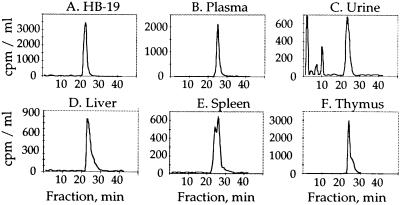
Figure 4.
HPLC radiochromatogram profile of various fluid and HB-19 target organs after a single injection of 20 μg (1.8 MBq) of [3H]-labeled HB-19—i.e., the plasma at 60 min (B), urine at 8–24 h (C), liver, thymus, and spleen (D–F; at 24 h postinjection). The HPLC profile of the native [3H]-labeled HB-19 is shown in A. [3H]-labeled peptide metabolites were revealed in the Sep-Pak extracted urine collected between 8 and 24 h postinjection. The slight stretching of the HB-19 peak in the liver, thymus, and spleen samples could be due to the partial association of HB-19 with its molecular partner in these tissues (see Fig. 5).

