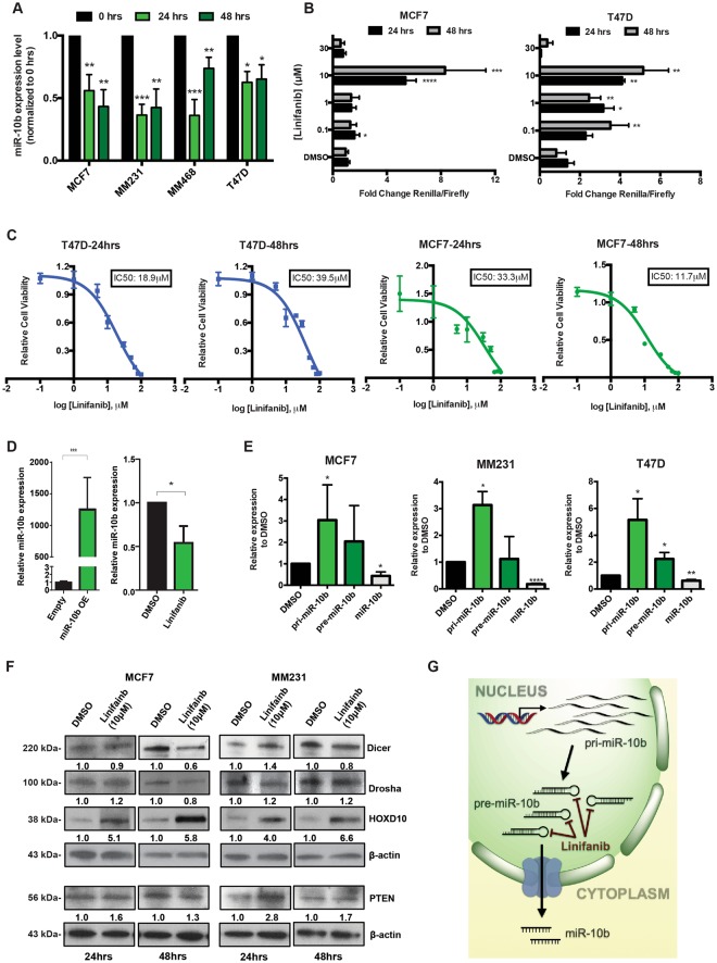
Figure 2.
Linifanib inhibits miR-10b expression in vitro in breast cancer cell line models. (A) Treatment with linifanib (10 µM) specifically decreases miR-10b expression levels in a panel of four breast cancer cell lines. (B) Dosage dependent assay was done in MCF7 and T47D cells, with the luciferase-based reporter construct after 24 and 48 hrs of treatment. (C) The IC50s of the compound at 10 µM where calculated via MTT assay after 24 (left panel) and 48 hrs (right panel) in T47D and MCF7 cell lines. The calculated IC50s are shown in boxes. (D) RT-qPCR analysis to confirm miR-10b overexpression in MCF7 clones compared to the empty vectors (left panel). miR-10b expression levels were determined in these MCF7 overexpressing clones by RT-qPCR after treatment with 10 μM linifanib. (E) RT-qPCR analysis to determine expression levels of primary-miR-10b (pri-miR-10b), precursor miR-10b (pre-miR-10b) and mature miR-10b after treatment with 10 μM linifanib for 24 hrs in breast cancer cell lines. (F) Western blots showing expression levels of Dicer, Drosha, HOXD10 and PTEN after treatment with 10 μM linifanib for 24 and 48 hrs in breast cancer cell lines. (G) Potential mechanism of action of linifanib: interaction with precursor miR-10b and reduction in the production of mature miR-10b with accumulation of primary and precursors transcripts of miR-10b. Error bars represent S.D. *represents P < 0.05, **represents P < 0.01, and ***represents P < 0.001.

